
Endoscopic Anatomical Swept-Source OCT: OCT systems are traditionally employed for high resolution imaging of tissues (such as measuring the thickness of retinal layers in opthalmology or examining the mucus lining of the respiratory epithelium) with a limited imaging range. In contrast, anatomical OCT systems have much longer imaging ranges and are therefore suitable for obtaining anatomical level details of organs and tissues.
Fig. 1. : Schematic of Endoscopic aOCT imaging of airway dynamics
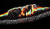
We have constructed anatomical OCT systems that utilizes a long flexible catheter to perform endoscopic imaging of the airways (Fig. 1) [Bu et al., 2017, Wijesundara et al., 2014]. The catheter is about 1.7m long and less than 1 mm in diameter; therefore, it can be introduced through the working channel of the smallest bronchoscope and allow imaging of the smallest bronchioles in the airway. Such imaging exams yield quantitative information about the airway shape and dynamics, enabling clinicians to better diagnose and treat airway diseases.
Our systems utilize a wavelength-swept laser source and an all fiber-optic interferometer. The sample arm of the interferometer consists of the endoscopic catheter that is connected to a stage that allows the catheter tip to be rotated and pulled back during imaging (Fig. 2). This allows the acquisition of either volumetric data of the inner surface of the airway as a helical scan or imaging at one position of the airway by using rotation alone as shown in Fig. 1. All components of the system are mounted on a portable cart that can either be transported to the patient's bedside or placed in a physician's office for an exam (Fig. 2).
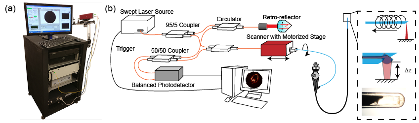
Fig. 2. : Swept-source anatomical OCT pediatric airways system; (a) the cart based aOCT system;
(b) block diagram of the system.
How is anatomical swept-source OCT different? In time-domain OCT, the back scattered signal from the sample is compared to the reflected reference signal. By changing the optical path length of the reference arm, the reflectivity and depth information of the layers within the sample is obtained and used to create images. Swept Source OCT is a type of Fourier Domain OCT in which instead of moving the reference, a special laser source produces a light beam whose wavelength sweeps over a range of different wavelengths at a very high rate. The wavelength sweep range determines the axial resolution of the system, while the instantaneous coherence length determines the imaging range. Anatomical Swept Source OCT systems utilize laser sources with a large sweep range and long instantaneous coherence length to obtain high-resolution images over an imaging range of the order of centimeters. This combination of features distinguishes anatomical swept-source OCT systems and enables imaging of large hollow organs within the body such as the airways.
intro page - research - publications - people - open positions
UNC Physics & Astronomy - Biomedical Research Imaging Center - UNC Home