
OCT for quantitative airway endoscopy: The airways are the portion of the respiratory system that transport air into the lungs; a narrowing of the airways can result in impaired breathing and can also result in life-threatening conditions in some cases. Airway obstruction can result from congenital or acquired deformations of the upper airways, such as subglottic stenosis. Airway narrowing can also be caused by dynamic collapse of the upper airway during sleep in conditions such as obstructive sleep apnea. Bronchoscopy is currently used to diagnose airway obstructions, it can provide a quick, real-time assessment of the airway anatomy, but yields only semi-quantitative results. Other methods of airway imaging, such as CT and MRI provide excellent quantitative information but involve exposure to ionizing radiation or lengthly examinations.

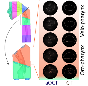
Fig. 1. : aOCT and CT images of a 3D printed pediatric airway of a 10 year old boy
Along with collaborators from the department of Otolaryngology/Head and Neck Surgery, we are developing a swept-source anatomical OCT (aOCT) system for airway imaging. This system can be used to perform quick and minimally-invasive assessments of airway obstructions, yielding quantitative, high-resolution information for improved diagnosis of obstructive airway disorders. The system can be used to produce real-time images of the dynamic airway morphology and also to produce 3D reconstructions of the airway anatomy [Bu et al., 2017, Wijesundara et al., 2014].
In order to evaluate the suitability of aOCT for airway imaging and to determine the optimal examination protocol, we have performed aOCT imaging in 3D printed models of the airway (Fig. 1), excised porcine tracheae and in vivo in mechanically ventilated pigs. We also routinely compare aOCT results with CT results to validate aOCT performance [Bu et al., 2017, Wijesundara et al., 2014, Balakrishnan et al., 2017]
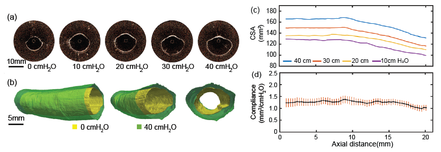
Airway elastography with aOCT: By combining changes in the airway geometry with respiratory pressure waveforms, we can derive estimates of the airway tissue mechanical properties (such as the tissue compliance, Fig. 2). Such measurements may be used to construct mathematical models of the airway and may be used to predict the likelihood of airway collapse [Bu et al, 2017]. Such models could also be used to model airflow changes occurring from surgical interventions and assist in the decision making process.

Fig. 2. aOCT derived compliance measurement in an ex vivo porcine trachea (a) Representative 2D slices under different applied pressures; (b) Comparison of 3D aOCT reconstructions at 0 cmH2O and 40 cmH2O; (c) Plots of cross sectional area under different pressures versus distance; (d) Varying Cross-sectional Compliance versus distance with standard error as the error bar.
intro page - research - publications - people - open positions
UNC Physics & Astronomy - Biomedical Research Imaging Center - UNC Home