
Magnetomotive Imaging with Superparamagnetic Iron Oxides: Superparamagnetic iron oxides (SPIOs) are used as a contrast agent for magnetomotive imaging in our lab. As their name suggests, SPIOs are tiny (generally less than 20 nm) particles which strongly exhibit paramagnetism – the magnetic dipole moments of their electrons’ spins cause them to align with an applied magnetic field. These particles are useful for biological imaging because, if administered to a patient or specimen, they can be controlled non-invasively by an externally-applied magnetic field.
The mechanism by which magnetomotive imaging works is outlined in the figure below. SPIOs are embedded in tissue or are attached to platelets in the bloodstream, as detailed in Pope et al, 2013. A magnetic field, produced by electromagnets, is applied over the imaging area, and when the field is modulated, the SPIOs feel a changing magnetic gradient force, dependent on particle volume, magnetization, and field strength. This force causes the SPIOs to oscillate up and down in phase with the changing magnetic field. As the particles move up and down, the tissue to which they are attached will also periodically deform slightly with the same frequency and phase. We use two different methods to detect this motion in our lab. Magnetomotive ultrasound imaging (MMUS) can be used to image several centimeters into tissue, while magnetomotive OCT (MMOCT) is used to image several millimeters deep, but at much higher resolution. In either case, we currently use frequency and phase-locking algorithms to determine which portions of the imaging area have undergone a mechanical phase shift, and conclude that such areas must contain SPIOs.
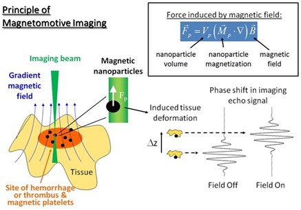
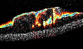
Fig. 1. Diagram detailing how magnetic particles are detected using interferometric techniques (from Oldenburg, et al, 2012).
The method for using OCT to detect magnetically labelled samples was first invented by Dr. Oldenburg as a postdoc in Dr. Stephen Boppart's laboratory in 2005 (Oldenburg et al, 2005). This technique was first used in vivo in 2006 to locate iron oxide particles in live tadpoles (Oldenburg et al, 2005). Since then, the technique has been further improved such that MMOCT can be used in more modern, spectral-domain OCT systems (Oldenburg et al, 2008). More recently, the Oldenburg lab demonstrated the ability of MMOCT to distinguish between healthy and injured pig arteries by attaching SPIO’s to platelets (Oldenburg et al, 2010). (Platelets are cells in the body which naturally move to wound sites in order to start the healing process.) For the most recent applications we are working on, please check out the thrombosis imaging page. Also, we have been using magnetomotive techniques for detecting magnetoreceptors in animals.
intro page - research - publications - people - open positions
UNC Physics & Astronomy - Biomedical Research Imaging Center - UNC Home